
TL;DR:
Getting the most from your AI screening report is largely in the imaging team’s hands. The report arrives finished, so it’s easy to read it as something the software decided on its own, but much of its accuracy is set at the table, in how each study is positioned, exposed, and collimated.
Here’s the veterinary AI report explained in short:
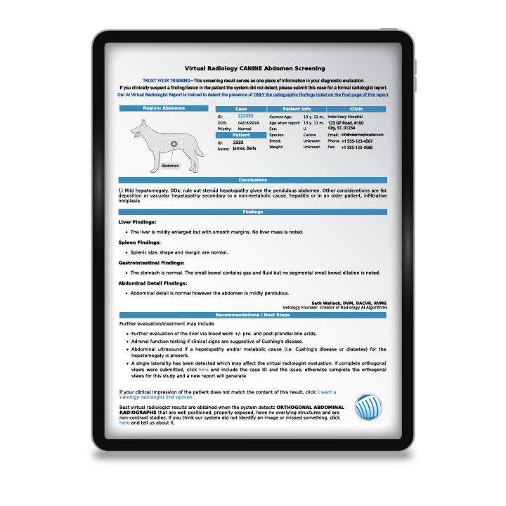
A header with study type, case ID, and signalment; a body region; a complete list of significant findings covering every visible organ, including the ones that look normal; and recommendations for next steps.
Because Vetology’s AI Report screens for more than 90 radiological changes, it may flag something unrelated to today’s presentation.
Five habits that improve every report:
- keep limbs out of the field of view
- reshoot images that are too dark or too light
- collimate tightly
- submit three views for thorax and abdomen (at least 2 for limbs)
- include complete anatomy with the landmarks visible
Reports arrive in five to seven minutes and open on any workstation, tablet, or phone. Read each one next to the radiograph, and let your clinical judgment guide the treatment plan. If a finding raises a question, the thumbs up or thumbs down button gets a Vetology radiologist to review the AI report within 24 hours at no charge. If the treatment plan still isn’t obvious, a full teleradiology report from a board-certified or board-eligible radiologist is available on demand (standard charges apply).
Veterinary medicine is a team sport, including imaging workflows. Clinics using Vetology’s AI screening and teleradiology services can make the most of each AI report by ensuring all team members work together to maximize image quality and understand exactly how the service works and what it needs to function optimally. AI is able to effectively accelerate and aid in the image interpretation process1 when the veterinary team improves their image inputs. The veterinary team then applies human knowledge to the report findings, allowing for more specific and targeted treatments.
Here’s our veterinary AI report explained.
What's in the AI screening report?
Learning how to read the AI radiology report from Vetology starts with knowing what to expect. At the top, you’ll see a header identifying the study type, the case ID, the patient’s signalment, client information, and your clinic’s details. Next to that, a body region diagram or a blue pill identifies the area under review.
Below that patient information, you’ll see a report with an easy-to-read and complete list of significant findings, including a breakdown of every condition classifier the AI reviewed in the study. If you submitted a thoracic or abdominal study, the AI will also look for changes in visible musculoskeletal structures. If multiple areas are imaged, the AI can generate either a comprehensive report or separate reports of each area (let us know which you prefer during install). For example, if thoracic and abdominal images are submitted, you can receive a musculoskeletal report showing any spinal abnormalities, a separate thorax report, and an abdominal report. Alternatively, you can receive a comprehensive report that combines findings from both regions. The report provides feedback on all visible organs, even those that appear clinically normal.

Because the AI screens for more than 90 radiological conditions or classifiers, it may point out issues in your patient that are unrelated to the current visit’s clinical signs, helping to ensure a more complete reading.
In most cases, the report will close with recommendations for next steps, which might include suggestions for additional imaging studies, other tests, or potential treatment options2 to consider for the suspected issue. If you need help understanding the AI’s recommended next steps, you can contact Vetology support for a quick response and to verify the AI’s findings.
Better images, better reports
Veterinary team members taking radiographs have a direct impact on the Vetology AI screening report’s accuracy.
“If the AI doesn’t have a good quality radiograph, it’s not going to be able to read the radiograph, or it’ll give false positives or false negatives,” says Vivian Paz, a veterinary technician with Vetology who reviews AI reports daily for accuracy. “Good positioning is a must.”
Here are some aspects of image quality Ms. Paz says team members can focus on to help clinicians get the most out of their AI screening reports:
- Positioning: A forelimb over the thorax can obscure the lungs or heart, and a hindlimb overlapping the abdomen can hide the bladder or other structures. Keep limbs out of the field of view.
- Exposure: Review your images – Underexposed or overexposed images can lead to inaccurate findings. If an image looks too dark or light, adjust your technique and reshoot it before submitting.
- Collimation: Tight, appropriate collimation improves image quality across the board.
- Views: A minimum of two views is necessary for the AI to interpret a given body area. Three is best for the abdomen and chest, and four or more is helpful for bone and MSK imaging (including obliques).
- Settings for small pets: The thorax and abdomen require different technique settings to get a good image, so whole-body images won’t give you good AI screening report results.
- Completeness: The Vetology AI screens only complete images, so you may get inferior or no results when the appropriate landmarks aren’t visible on the film.
- For very large dogs whose bodies require multiple shots per body area, please ensure organs in your images overlap each other and include complete organs. You can also jump directly to interpretation by a board-certified or board-eligible radiologist.
Read the AI report alongside your images
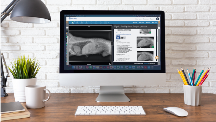
The report is meant to be read alongside the radiograph, not instead of it. Pull the images up beside the report on our platform and work through them together. The AI screens for patterns across every visible structure; you hold the patient’s history, the physical exam, and the rest of the clinical picture that decides what the combination of each single finding actually means. When the report and your read agree, that’s a useful confirmation. When they don’t, that’s worth something too. Either way, your medical expertise guides the treatment plan.
If something in the report raises a question, you don’t have to resolve it alone. Every case has a thumbs up and thumbs down feedback button associated with it. Click it, and our imaging techs will review it with a Vetology radiologist and get back to you within 24 hours on weekdays. That review is always free and encouraged. It isn’t a full teleradiology report, and it isn’t meant to stand in for one; it’s a straight answer about what the AI flagged and why. When a case calls for an independent specialist interpretation, order a full teleradiology report at our standard fees.
The in-platform support chat, phone and email are all staffed by people, not bots. They can help with questions about reports, your images, technical troubleshooting, or anything else related to our service.
You don’t need to be an AI expert to get the most out of your AI reports. You just need to have some on speed dial, and that’s where our support team shines.
AI screening report workflows
Once your clinic’s DICOM connection is set up with Vetology, every set of radiographs you take goes straight to the platform without the need to upload images separately.
The AI generates a screening report within five to seven minutes, and because the platform is web-based, anyone on the team can pull up the report from any workstation, tablet, or phone. You don’t need to be in the X-ray room to access results.
If the AI detects something that doesn’t quite make sense when considering the clinical picture, or if the case is complex enough to warrant a specialist opinion, the veterinarian can choose to escalate the case to a boarded (or board-eligible) veterinary radiologist through the Vetology platform. This system supports veterinarians and is in keeping with the American College of Veterinary Radiology (ACVR) and the European College of Veterinary Diagnostic Imaging’s position on how AI tools should enhance, not impede,3 a veterinary professional’s diagnosis and treatment.
Using the report as a team
If there’s one thing to take from this guide, it’s that the quality of an AI report is decided long before the report exists. It starts at the table, in the way each study is positioned, exposed, and set up. That’s good news, because much of the outcome is in the imaging team’s hands. Making the most of AI in radiology requires developing a high-quality workflow.4 Training your team in this mindset is important for successful use of AI veterinary radiology screening tools.
A clean, well-positioned study gives the AI its best chance to find what matters, including early changes unrelated to the patient’s presentation. That kind of catch starts with the technician in the X-ray suite. So keep the images clean, learn what the AI can flag, and read each report along with the radiograph. It’s how a routine radiograph becomes a fuller picture of the patient’s health, and how the report earns its place as a fast, reliable second look for every case.
References
1Lim, Sungwon. “AI helps your veterinarian provide optimal care for your dog or cat.” AnimalWellnessMagazine.com. August 21, 2022. Accessed August 26, 2026. https://animalwellnessmagazine.com/ai-helps-your-veterinarian-provide-optimal-care-for-your-dog-or-cat/.
2Ontario Veterinary College. “Harnessing the Power of Artificial Intelligence in Veterinary Medicine.” UOGuelph.ca. December 4, 2023. Accessed August 26, 2026. https://www.uoguelph.ca/ovc/news/node/2628.
3Appleby, Ryan B., Difazio, Matthew, Cassel, Nicolette, Hennessey, Ryan, Basran, Parminder S. “American College of Veterinary Radiology and European College of Veterinary Diagnostic Imaging position statement on artificial intelligence.” AVMAJournals.avma.org. March 19, 2025. Accessed August 26, 2026. https://avmajournals.avma.org/view/journals/javma/263/6/javma.25.01.0027.xml.
4Jahagirdar, Madhu. “Transforming the Radiology Experience Through Integrated AI.” ITNOnline.com. August 17, 2026. Accessed August 26, 2026. https://www.itnonline.com/article/transforming-radiology-experience-through-integrated-ai.