
Behind The Scenes With the Vetology Support Team
In veterinary medicine, time is short and expectations are high. Clients want answers about their pet’s health quickly, and AI-powered platforms like Vetology can help you deliver. But what happens when you have a question about your AI screening report, need to speak with a human radiologist, or want to train your team to use the platform?
Our client care team is ready to help at a moment’s notice. Clients who regularly interact with the support team tend to get better results from the platform, work more efficiently, and build greater confidence with our AI and teleradiology tools.
Here’s a look at the Vetology support services we provide at no additional cost to help users get the most from our platform.
The Team Behind The Screen
Vetology’s support team is small but mighty. Together, they handle over 14,000 communications each year, including phone calls, emails, scheduled trainings, and now, live on-platform chats.
Our support providers include a blend of veterinary techs, technology, and customer care professionals. With many years of combined experience across multiple disciplines, they’re capable of handling everything from onboarding and software installation to troubleshooting, clinical questions, radiologist follow-ups, and veterinary team coaching.
We’d like to introduce you to two of our key support team members:
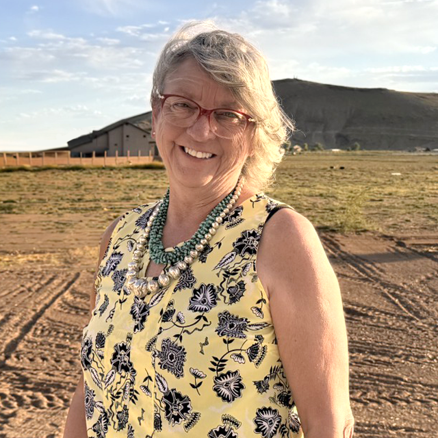
Tammie McGill
Tammie McGill spent nearly two decades as a human EMT before transitioning into a role as a veterinary assistant. After gaining years of clinical experience, she now uses her strong veterinary technician skills to provide clinical support to Vetology users, which includes answering AI report questions, coordinating discussions with interpreting veterinary radiologists, monitoring radiograph quality, and helping clinical teams troubleshoot imaging techniques to improve safety and optimize outcomes.
Tammie and her fellow veterinary technician, Vivian Paz, also work closely with the radiologists, data scientists, and development teams, offering valuable advice and domain-specific insight.

Sandra Nemis
Sandra Nemis came to Vetology after several years of managing customer care teams, including a technical supervisor role.
She now leads the Vetology support team through client interactions, handles clinic demos, installations, onboarding, training, and day-to-day platform support.
With the help of additional support team members, Aziz Beguliev, Chey Aranzasu, Kath Dato, and our SVP of Information Systems Ruben Venegas, Tammie and Sandra ensure that no question goes unanswered and no case falls through the cracks.
While most have been on the team for more than five years, tenures span from new members to 15 years, reflecting a mix of institutional knowledge and fresh perspectives to help deliver consistently excellent service and fast communication.
From Demo to Diagnostics
When clinics reach out to Vetology through the website, email, or phone, they establish a relationship with our tight-knit client care team from day one.
The onboarding process for new Vetology clients is quick and efficient. After a client completes a short form with clinic information, the Vetology support team creates their internal profile, configures platform access, and schedules an installation and training session.
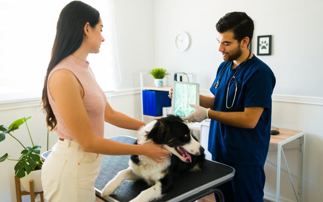
“We remote into the X-ray computer, add our destination settings to ensure communication, and enable the auto-send feature,” explained Sandra. “When team members take X-rays, they don’t have to do anything extra; the images automatically go to the Vetology platform. Within a few minutes, they have an AI screening report and can submit to a board-certified radiologist, if desired.”
The entire process of installing and configuring the platform and providing initial training to key team members typically takes less than an hour, so you can be up and running fast and avoid downtime in the clinic.
Clinical Coaching and Aftercare
Vetology’s support combines technical help with clinical collaboration. Our two veterinary support specialists have nearly three decades of combined experience. Together, they provide a crucial “aftercare” service for teams using the Vetology platform.
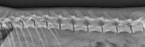
When the team spots an issue with image quality or safety, they provide feedback and coaching. They can offer tips for technicians to hone their radiology skills and how to use positioning aids, something that they may or may not have learned or practiced in school.
“Clinics are very responsive when we reach out,” said Tammie. “I’ve also had doctors call to ask, ‘What else can we do to make this better?’ I’ll talk to anybody in the clinic that has time or is willing to learn more.”
Coaching support helps improve image quality and diagnostic accuracy, reduce retakes, and protect team members from unnecessary radiation exposure.
Contacting Vetology Support
You can contact Vetology’s support team by phone, email, the website, or the live chat feature on the platform.
However you choose to contact the team, you can expect a rapid response. The team is available from 6 a.m. to 6 p.m. Pacific time, and responds to emails during regular hours within five to 10 minutes. If you have a question after hours, send an email you can expect a response first thing the following morning.
Most importantly, when you contact Vetology’s support team, your questions will be answered by a real person. Our goal is to provide quick help so you get the most from our platform without slowing down your day.
Practice Support That Delivers
The best veterinary technology platforms and imaging tools not only provide a place to process images, but they also help teams use them to their full extent. Vetology’s support team aims to provide accessible, proactive help during your daily workflows, when you need it most. We want to ensure that clinics feel supported, confident, and ready to make the most of every feature.
When clinics use our responsive support, teams learn to optimize their images and submissions, radiologists and AI screenings have higher-quality studies to work from, reports become more accurate, and pets receive better, more timely care.
Trusted Support is a Click or Call Away
Our helpful, professional, human support team knows your clinic, understands your challenges, and wants you to succeed. From onboarding to aftercare, we’re committed to helping clients use our AI and teleradiology systems more confidently every day.