Guest Post: The Power of AI in Veterinary Medicine
At Avoca Drive Animal Hospital, the integration of Vetology’s AI tools with their Practice Management Software has been a collaborative effort focused on supporting veterinary teams and improving clinical efficiency. This partnership has helped streamline workflows, reduce daily workload, and free up valuable time for patient care. By working closely with Vetology’s team, Avoca Drive has created a more seamless diagnostic process—enhancing clinical confidence and decision-making when it matters most. In this article, you’ll learn how thoughtful AI integration and strong ongoing support have made a measurable impact in a busy veterinary practice. In this article you will learn how:
- AI in veterinary medicine integrates directly into practice management software.
- Vetology’s AI reduces manual tasks, giving vets more time for patient care.
- AI reports support clinical judgment, enhancing diagnostic confidence.
- In emergencies, AI in veterinary medicine helps vets make faster more informed decisions.
- Combining AI with clinical expertise leads to better outcomes and a higher standard of veterinary care.
The Power of AI in Veterinary Imaging:
Revolutionizing Diagnosis and Treatment
Written by: Dr Elizabeth O’Connor BVSc, FFCP, Veterinarian, CEO, Author
In the ever-evolving field of veterinary medicine, technology plays a crucial role in improving patient care. At our veterinary hospital, we have embraced AI technology—specifically Vetology—for imaging, and the benefits have been remarkable. Let’s explore how this innovative tool has transformed our diagnostic processes and enhanced treatment decisions, ultimately leading to better outcomes for our furry patients.

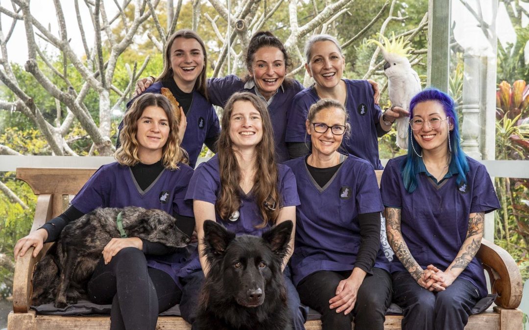
Meet the team at Avoca Drive Animal Hospital, a Fear Free certified Practice.
Streamlining Workflow for Efficiency
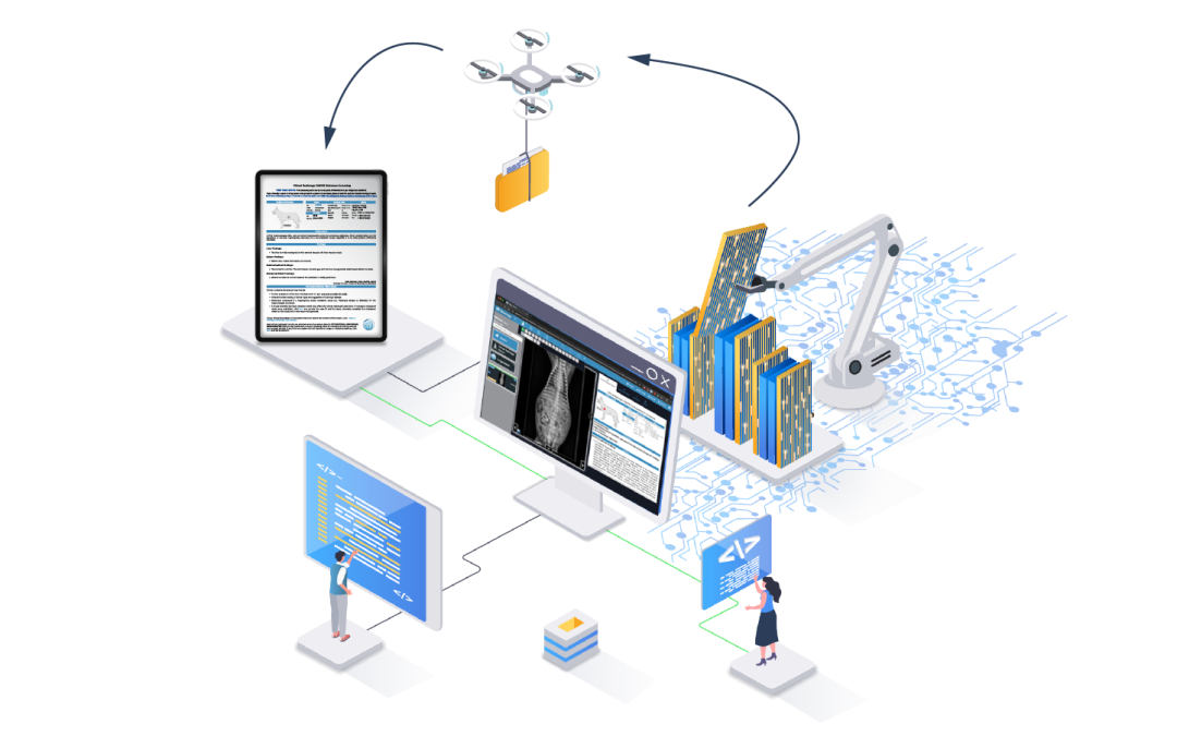
From the moment we integrated Vetology into our practice, we focused on making the transition as seamless as possible. Collaborating with our Daysmart software team, we completed the integration within just a couple of days. This investment in time and effort has proven invaluable.
Now, AI-generated reports flow directly into our practice management software, as well as the Vetology workstation platform. This integration eliminates the cumbersome process of manually uploading and downloading files, which previously consumed precious time that our team needed to dedicate to patients and clients. By streamlining our workflow, we can focus more on providing exceptional care.
Enhanced Diagnostic Confidence
Once AI results are incorporated into a patient’s file, our veterinarians leverage this data alongside their expertise. Each case prompts our team to critically analyze the findings:
“Does this make sense given the patient’s history, signalment, and other information that the AI doesn’t have?”
With a comprehensive view that includes history, blood results, and AI insights, our veterinarians gain a clearer understanding of the patient’s condition. This is especially crucial in urgent cases, such as those involving abdominal pain or suspected gastrointestinal issues.
The added assurance that AI provides helps our team confirm that nothing has been overlooked, reducing stress during busy times when multiple cases demand attention.
Making Timely Decisions for Critical Care
When our vets have a clear plan of action, informed by both their clinical judgment and AI reports, we are much better positioned to act swiftly. In urgent situations, this can mean the difference between life and death for seriously ill patients. Rather than waiting for specialist opinions or navigating uncertainties, we can initiate the necessary procedures immediately.
This rapid response allows us to explain diagnoses and obtain consent from pet owners much more efficiently. When every second counts, having reliable AI insights empowers our team to make informed decisions and provide the timely care that pets need.
Conclusion: A New Era in Veterinary Medicine
The integration of AI technology in our veterinary hospital has transformed the way we approach diagnostics and treatment. By streamlining our processes, enhancing diagnostic confidence, and facilitating timely decision-making, we are better equipped to provide exceptional care for our patients.
As we continue to embrace innovative technologies like Vetology, we remain committed to improving the health and well-being of the pets we serve. The future of veterinary medicine is bright, and we are excited to lead the way in providing the highest standard of care.