Another pair of Eyes: 4 Reasons Veterinary Teleradiology is a Smart Move (Even If You’re an Expert)

Another pair of Eyes: 4 Reasons Veterinary Teleradiology is a Smart Move (Even If You’re an Expert)

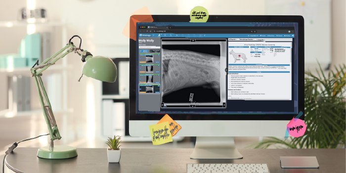
Interpreting radiographs and other diagnostic images is a core skill for every veterinarian. Still, even the most experienced practitioners know that an extra layer of review can make or break the outcome of a case. Whether you’re a solo practitioner, managing an overnight emergency, or covering cases while the radiologist is on leave, veterinary teleradiology can provide the clarity and support you need to make confident treatment decisions.

Vetology connects clinicians with board-certified radiologists who specialize in interpreting complex diagnostic images. You might think of veterinary teleradiology as merely a backup option for less experienced practitioners. However, it can be a valuable tool that encourages multidisciplinary collaboration among veterinarians in diverse practice areas and career stages. Here are four reasons teleradiology is meant for veterinarians of all skill levels.

1. Collaborative Medicine is Good Medicine

In human healthcare and at veterinary specialty hospitals, surgeons, internists, and radiologists routinely consult with one another before proceeding with treatment. Veterinary teleradiology extends that same collaborative model to veterinary general practice hospitals and urgent care clinics, supporting informed decisions in everyday care.

Opting for a teleradiology consult can feel like you’re second-guessing yourself, but it’s actually a powerful way to strengthen your clinical instincts. Working with veterinary radiologists can enhance your interpretation skills and reinforce your decision-making. Veterinary radiologists can spot subtle signs in radiographs, dental radiographs, ultrasounds, and CT scans that, left unnoticed, can drastically alter the course of treatment.

2. Faster Answers Mean Better Outcomes

Another significant advantage of veterinary teleradiology is availability and speed. When an in-house radiologist is unavailable and the case is urgent, waiting may not be an option. Teleradiology provides your practice with 24/7 access to expert readings, offering a fast turnaround that enables timely treatment.

Rapid answers are especially critical when dealing with emergency conditions, such as:

  • Gastrointestinal obstruction
  • Heart failure
  • Pneumonia
  • Pneumothorax

“Anytime there’s a time factor on a critical case, that’s where telemedicine is at its best,” said Dr. John Mattoon, board-certified veterinary radiologist and senior medical advisor for Vetology.

GI cases are among the most challenging diagnoses that general practitioners face regularly. Teleradiology doesn’t replace the expert opinion, skill, or clinical instincts of the veterinarian, but it can help you prevent serious misdiagnoses.

“Looking at the bowel of vomiting dogs and cats is the most challenging thing we do,” said Dr. Mattoon. “For a veterinarian to say, ‘Your dog is obstructed, you need to go to surgery’ is a pretty bold move. You’d better be right.”

A few other conditions Dr. Mattoon noted as difficult to diagnose without an expert eye included gallstones, which can appear superimposed over the liver on lateral views, and gas in the hepatobiliary system, a subtle change that can provide insight into why a pet is so sick. “Gas within the bile ducts, gallbladder, or the liver itself is not obvious, but when it’s there, it indicates an anaerobic infection or abscess. If you miss it, that condition is often fatal.”

3. Expert Interpretation Supports Skill Development

There’s growing concern that some veterinary teams, especially new graduates, rely too heavily on teleradiology and that overreliance can undermine clinical development and decision-making skills. However, used appropriately, veterinary teleradiology can help early-career veterinarians develop and reinforce those skills.

Radiology reports can be an incredible learning tool. “The opportunity is tremendous for self-learning,” Dr. Mattoon noted. “You have a detailed report that explains the abnormalities on the image, followed by recommendations for appropriate next steps.”

When you commit to studying the images and reports, you can learn and grow with each case. Technicians and support staff can benefit, too. If the radiologist includes their contact information with the image report, you can call with questions about the case to further your understanding of the pathology.

4. Teleradiology Meets A Growing Need

Veterinary radiologists are in short supply, particularly in academia, which limits the training of new specialists. In areas without access to a local radiologist, primary care veterinarians must find ways to meet client and patient care needs in-house, including the use of veterinary teleradiology. “Teleradiology has been huge in allowing DVMs to access radiologists nearly instantaneously,” said Dr. Mattoon. “We can serve more veterinarians and do so more efficiently.”

Teleradiology can help general practitioners with the following scenarios that expand the scope of primary care:

  • Second opinions when another clinician is unavailable
  • Serious, urgent cases with no room for error
  • Client specialist consultation requests
  • Abdominal and thoracic ultrasound interpretation

Teleradiology For All

Veterinary teleradiology can benefit everyone, from new graduates and solo practitioners to experienced clinicians, emergency veterinarians, and even boarded specialists. Any veterinarian who values accuracy, collaboration, and providing the highest standards of patient care can benefit from image interpretation services such as the teleradiology service offered by Vetology.

Contact us for help navigating your next challenging case or when you need a second set of eyes and a fresh perspective to enhance patient care and client service in your clinic. Vetology’s teleradiology service is hassle-free, contract-free and allows flexibility with a pay as you go model. Our team of boarded radiologists, a boarded dental specialist and a cardiologist offer industry-standard STAT and turnaround times, and are available to read canine, feline, equine, exotics, avian and reptile cases.

AI and Teleradiology Questions: Answered

To learn more about Vetology and see our platform in action, click this box, to contact the Vetology support team.

Is AI Better Than a Veterinary Radiologist at Reading Pet X-rays?

Is AI Better Than a Veterinary Radiologist at Reading Pet X-rays?

This article examines the comparison between using AI in veterinary radiology and the human experience. Even though AI does improve efficiency by pre-screening X-rays and generating reports, it cannot replace radiologists due to variability in interpretation. AI performs best in clear conditions with strong expert agreement, while complex cases still require human expertise. Read more about how AI in radiology:

  • Addresses the shortage of veterinary radiologists.
  • Helps with pre-screening and structured reports.
  • Works well for conditions like hepatomegaly or pericardial effusion.
  • Supports, not replaces, veterinary radiologists.

AI Versus Veterinary Radiologists: Collaboration, Not Competition

About 94 million U.S. households own at least one pet.[1] That’s a lot of furry, feathered, and scaly family members that may potentially need radiographs to diagnose a medical condition. However, there are only 667 board-certified radiologists in the country [2] creating a bottleneck in radiology services. This shortage can correlate to longer wait times, increased anxiety for clinicians and pet owners, and potential delays in diagnosing critical conditions.

This is where artificial intelligence-based radiology tools can help—not to replace veterinary radiologists, but to support them. Artificial intelligence (AI) can pre-screen images, highlight abnormalities, and generate structured reports, allowing radiologists to focus on complicated cases while improving efficiency for general practitioners. But, how does AI compare to human expertise?

Not all conditions are created equal

Radiology is not an exact science but rather an interpretive discipline that relies on pattern recognition, clinical judgement, and experience. Board-certified veterinary radiologists undergo extensive training, but they don’t always agree on image interpretations, especially if the changes are subtle or the patient has multiple diagnoses, creating overlapping signs.

Studies have shown that radiologists tend to have a high level of agreement when interpreting X-rays that display clear and advanced disease. However, variability in interpretation increases when findings are more subtle, as may be the case in early-stage tumors, mild joint changes, or diffuse lung patterns that could indicate interstitial or early inflammatory disease. When subtle abnormalities are suspected, additional imaging, such as ultrasound, computed tomography (CT), or magnetic resonance imaging (MRI), can provide greater anatomical detail and diagnostic confidence.

How interpretive variability affects AI performance assessment

Understanding variabilities in radiologist interpretations is necessary to fairly evaluate the AI’s diagnostic accuracy, sensitivity, and specificity.

  • AI algorithms rely on human-labeled data (i.e., ground truth) to learn how to detect and classify abnormalities, and if radiologists don’t agree on a diagnosis, the ground truth may have some degree of subjectivity.
  • AI radiology tools are evaluated using accuracy, sensitivity, and specificity, but these measures must be analyzed in the context of how consistently radiologists themselves diagnose the condition.
  • If two radiologists interpret the same case differently, the AI may match one but disagree with the other. This doesn’t mean that the AI is wrong; it only highlights the inherent variability in radiology.

How interpretive variability affects AI radiology use

The inherent variability in veterinary radiology associated with certain conditions means that some are well-suited for AI screening while others aren’t.

For example, conditions such as hepatomegaly, esophageal enlargement, and the presence of pericardial effusion have a high radiologist agreement rate and are well-suited for AI screening.

At Vetology, each AI-generated report includes a clear list of the conditions assessed, so it’s clear exactly what was evaluated, what was flagged, and what falls outside the scope of the current screening. This provides veterinarians with a solid understanding of the AI’s capabilities and limitations, enabling them to focus their clinical decisions on conditions that were not screened for, without expecting input on findings beyond the AI’s parameters.

image of Vetology's AI report featured on a tablet or ipad

Vetology’s AI tools provide guidance for a wide range of thoracic, abdominal, and musculoskeletal conditions in canine and feline patients, including—but not limited to—the following

Abdominal Classifiers

  • Liver enlargement
  • Masses that may indicate neoplasia or inflammatory processes
  • Splenic changes, commonly linked to systemic or localized disease
  • Kidney abnormalities such as mineral deposits, structural size variations that may suggest neoplasia, inflammation, or systemic disease
  • Bladder and urethral stones
  • Pregnancy detection
  • Gastrointestinal tract abnormalities, which may indicate obstruction, motility issues, or other conditions
  • Peritoneal fluid accumulation, inflammation, or infection

Thoracic classifiers

  • Pulmonary patterns
  • Cardiomegaly
  • Pleural fissure lines
  • Fluid accumulation
  • Soft tissue pulmonary nodules
  • Masses
  • Vascular enlargement

Leveraging AI screening alongside teleradiology

Vetology allows veterinarians to optimize AI radiology screening tools and teleradiology services to enhance diagnostic accuracy, improve efficiency, and expedite patient care.

For example, let’s say you handle 60 X-ray cases a month, and you send out only 10 for teleradiologist review to avoid the expense. A Vetology subscription, which provides unlimited access to AI screening and full reports in as little as five minutes, could support your clinical expertise, helping to confirm your suspicions and streamline decision-making. If you still have doubts about a case, you can escalate it for review by a board-certified veterinary radiologist.

This approach creates a three-tiered approach to patient care, integrating:
• AI insights
• With your professional judgement,
• and expert validation from a radiologist when needed.

Collaborating with the Vetology team can help ensure that your patients receive a timely diagnosis and treatment plan, allowing them to receive the care they deserve quickly.

radiograph showing a well positioned and collimated Canine Thorax

How you can support accurate AI screening and faster board certified radiologist reports

One of the most important factors that lead to an accurate AI screening is good radiographic technique. Clear, well-positioned, well-developed radiographs are necessary for accurate human and AI interpretation, and the AI does not have the ability to adjust its interpretation based on altered positioning or an unclear image.

For example, if a patient is slightly twisted, anatomical structures may appear distorted on the image. This can lead the AI to misread the size or shape of an organ, or even misidentify a condition. Human radiologists can identify when a patient isn’t perfectly positioned and adjust their interpretation, but AI doesn’t yet have that context—it reads exactly what’s in front of it.

You can take the following measures to increase the likelihood of accurate AI screening:

  • Ensure proper positioning of each patient
  • Choose the correct radiographic settings to ensure a clear image
  • Take at least two views (ventrodorsal and lateral views) of the area to be assessed every time.
  • Collimate down to the region of interest to reduce scatter.

Vetology offers personalized, on-demand support tailored to answer your needs and questions. Our team of radiologists and veterinary technicians is always available to provide free, one-on-one guidance with positioning skills and technical assistance (in some cases), whether you’re a seasoned practitioner, a new team member, or a recent graduate.

References
[1] According to the American Pet Products Association (APPA) 2025 State of the Industry Report published stats in Today’s Veterinary Business, April, 2025.
[2] AVMA published statistics – veterinary specialists in the United States as of December 31, 2024.AVMA published statistics – veterinary specialists in the United States as of December 31, 2024.

AI and Teleradiology Questions: Answered

To learn more about Vetology and see our platform in action, click this box, to contact the Vetology support team.

Veterinary Radiology AI: Ensuring Accuracy, Trust, and Quality Care

Veterinary Radiology AI: Ensuring Accuracy, Trust, and Quality Care

This article discusses how Vetology’s Radiology AI Tool was created to improve diagnostic accuracy, streamline workflows, and support veterinarians with reliable first-line screening. The system leverages advanced AI methods like CNNs, QA testing, and LLMs for report generation. Designed as a supportive tool, it enhances trust and care quality without replacing expert radiologists. Learn how AI supports veterinary radiology by:

  • It is built on a large dataset labeled by board-certified radiologists.
  • Using CNNs, confusion matrices, QA testing, and LLMs.
  • Improving consistency with preprocessing, cropping, and anomaly detection.
  • Undergoing continuous updates with clinical data and structured reviews.
  • Enhancing workflow efficiency while leaving final judgment to experts.

When you take a radiograph to better understand a patient’s condition, an accurate reading of the image is key to ensuring the animal receives appropriate treatment. That’s why U.S. board-certified radiologists on the Vetology team work with our data scientists and developers to hone our artificial intelligence (AI) models. By integrating a diverse team of subject matter experts, and combining their skills with rigorous testing, and quality assurance measures, AI can support diagnostic efficiency while maintaining the trust and reliability veterinarians need for patient care.

To help you better understand the Vetology AI radiology tool, this article explains how it was developed, validated, and how we iterate on our models.

Relying on the Experts

​To develop our AI model, we used more than a million images from hundreds of thousands of cases, ensuring a comprehensive representation of anatomical variations and disease conditions. Each image was evaluated and annotated by a U.S. board-certified veterinary radiologist, providing high-quality, expert-labeled data (i.e., ground truth) that allows the AI to learn from professional interpretations.

Training the AI

To train our veterinary radiology AI tool, we used a combination of deep learning techniques, including convolutional neural networks (CNNs), confusion matrices, quality assurance (QA) regression testing, and large language models (LMMs).

Convolutional Neural Networks

CNNs are designed for image recognition and pattern detection, enabling the automated analysis of radiographs with high accuracy. The images first undergo preprocessing to ensure consistency. This includes image orientation, maximizing image clarity, and contrast adjustments. The CNN then learns to identify features and detect patterns.

  • The first convolutional layers identify edges, textures, and contrasts, distinguishing bones, organs, and soft tissues.
  • Multi-output CNNs can determine whether an X-ray belongs to a dog or cat and pinpoint the anatomical region being analyzed.
  • Once trained, a CNN can determine orientation and recognize certain abnormalities and conditions.

Confusion Matrices

A confusion matrix helps measure how well an AI model classifies radiographic images, ensuring it can correctly identify normal versus abnormal scans, specific conditions, and disease severity. It compares the AI’s predictions with the ground truth, which is determined by U.S. board-certified veterinary radiologists. The table below outlines the relationship between the four key components:

chart showing the different outcomes for a confusion matrix

When used to evaluate results, the confusion matrix describes the AI’s performance by measuring key performance metrics, including:

  • Accuracy = (TP + TN) / total cases
  • Sensitivity = TP / (TP + FN) — How well the AI detects conditions
  • Specificity = TN / (TN + FN) — How well the AI identifies normal cases
  • Precision = TP / (TP + FP) — How many positive predictions are correct
  • F1 score = The balance between precision and recall, ensuring AI does not over or under diagnose

Quality Assurance Regression Testing

QA regression testing compares AI-generated results with known labeled images to identify errors, inconsistencies, and areas for improvement. This process enables our developers to fine-tune the AI, reducing false positives and false negatives, and thus enhancing results over time.

Large Language Models

Large Language Models (LLMs) are trained to recognize and generate common veterinary diagnostic phrases, sentence structures, and condition descriptions to create professional and structured reports.

Board-certified veterinary radiologists are once again involved to review the generated phrases and confirm that the AI is accurately interpreting the images and the LLM is producing relevant and coherent statements.

AI Screening Features

AI screening features enhance veterinary radiology through efficiency tools that promote improved AI reports and consistent image interpretation. Key features include:

  • Image preprocessing and standardization: Pre-AI tools adjust orientation, brightness, and contrast for clearer analysis.
  • Automated cropping: EfficientDet SSD technology isolates the area of concern, improving contextual accuracy for AI interpretations.
  • Anomaly detection: AI identifies abnormalities, such as fractures, tumors, and changes in lung patterns, and can detect species- and region-specific changes. Severity grading models can also help classify the condition’s severity.

Keeping Updated

To ensure our AI model remains accurate and aligned with evolving veterinary radiology practices, we regularly update it with new data to integrate the latest medical findings and maintain optimal performance. The modifications undergo a structured change management process to ensure the updates improve accuracy without introducing errors, and we track all changes between AI versions to maintain transparency and traceability of updates.

Vetology’s AI radiology model is designed to support, not replace veterinary expertise, improving image analysis and providing clinicians with faster, more consistent insights. Utilizing an AI radiology tool can help veterinary teams make more informed decisions before seeking expert consultation. Veterinarians can use this tool as an initial screening step before sending cases to a teleradiologist, helping streamline workflows, prioritize urgent cases, and improve diagnostic efficiency.

Want to see AI in action?

To learn more, contact our Vetology team, or book a demo for a firsthand look at our AI and teleradiology platform.

Pin It on Pinterest