Veterinary Radiology AI: Ensuring Accuracy, Trust, and Quality Care

Veterinary Radiology AI: Ensuring Accuracy, Trust, and Quality Care

When you take a radiograph to better understand a patient’s condition, an accurate reading of the image is paramount to ensure the animal receives the appropriate treatment. That’s why U.S. board-certified radiologists on the Vetology team worked in conjunction with the technology crew to hone our artificial intelligence (AI) models. By integrating expert oversight, rigorous testing, and quality assurance measures, AI can support diagnostic efficiency while maintaining the trust and reliability veterinarians need for patient care.

To help you better understand the Vetology AI radiology tool, this article explains how it was developed and validated, and how it is improved.

Relying on the Experts

​To develop our AI model, we used more than a million images from hundreds of thousands of cases, ensuring a comprehensive representation of anatomical variations and disease conditions. Each image was evaluated and annotated by a U.S. board-certified veterinary radiologist, providing high-quality, expert-labeled data (i.e., ground truth) that allows the AI to learn from professional interpretations.

Training the AI

To train our veterinary radiology AI tool, we used a combination of deep learning techniques, including convolutional neural networks (CNNs), confusion matrices, quality assurance (QA) regression testing, and large language models (LMMs).

Convolutional Neural Networks

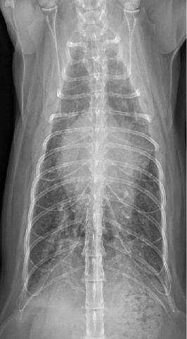
CNNs are designed for image recognition and pattern detection, enabling the automated analysis of radiographs with high accuracy. The images first undergo preprocessing to ensure consistency. This includes image orientation, maximizing image clarity, and contrast adjustments. The CNN then learns to identify features and detect patterns.

  • The first convolutional layers identify edges, textures, and contrasts, distinguishing bones, organs, and soft tissues.
  • Multi-output CNNs can determine whether an X-ray belongs to a dog or cat and pinpoint the anatomical region being analyzed.
  • Once trained, a CNN can determine orientation and recognize certain abnormalities and conditions.

Confusion Matrices

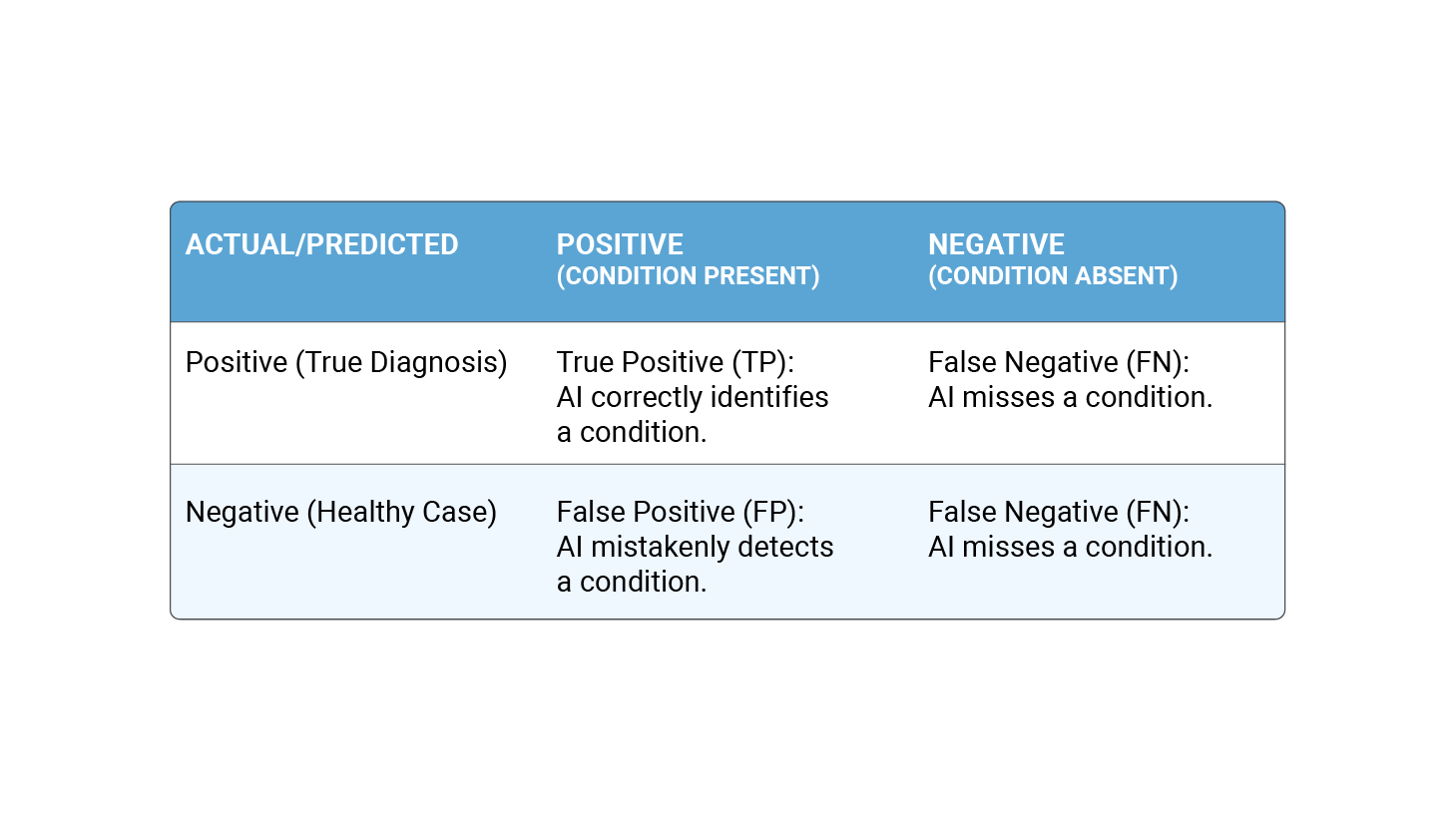
A confusion matrix helps measure how well an AI model classifies radiographic images, ensuring it can correctly identify normal versus abnormal scans, specific conditions, and disease severity. It compares the AI’s predictions with the ground truth, which is determined by U.S. board-certified veterinary radiologists. The table below outlines the relationship between the four key components:

chart showing the different outcomes for a confusion matrix

When used to evaluate results, the confusion matrix describes the AI’s performance by measuring key performance metrics, including:

  • Accuracy = (TP + TN) / total cases
  • Sensitivity = TP / (TP + FN) — How well the AI detects conditions
  • Specificity = TN / (TN + FN) — How well the AI identifies normal cases
  • Precision = TP / (TP + FP) — How many positive predictions are correct
  • F1 score = The balance between precision and recall, ensuring AI does not over or under diagnose

Quality Assurance Regression Testing

QA regression testing compares AI-generated results with known labeled images to identify errors, inconsistencies, and areas for improvement. This process enables our developers to fine-tune the AI, reducing false positives and false negatives, and thus enhancing results over time.

Large Language Models

Large Language Models (LLMs) are trained to recognize and generate common veterinary diagnostic phrases, sentence structures, and condition descriptions to create professional and structured reports.

Board-certified veterinary radiologists are once again involved to review the generated phrases and confirm that the AI is accurately interpreting the images and the LLM is producing relevant and coherent statements.

AI Screening Features

AI screening features enhance veterinary radiology through efficiency tools that promote improved AI reports and consistent image interpretation. Key features include:

  • Image preprocessing and standardization: Pre-AI tools adjust orientation, brightness, and contrast for clearer analysis.
  • Automated cropping: EfficientDet SSD technology isolates the area of concern, improving contextual accuracy for AI interpretations.
  • Anomaly detection: AI identifies abnormalities, such as fractures, tumors, and changes in lung patterns, and can detect species- and region-specific changes. Severity grading models can also help classify the condition’s severity.

Keeping Updated

To ensure our AI model remains accurate and aligned with evolving veterinary radiology practices, we regularly update it with new data to integrate the latest medical findings and maintain optimal performance. The modifications undergo a structured change management process to ensure the updates improve accuracy without introducing errors, and we track all changes between AI versions to maintain transparency and traceability of updates.

Vetology’s AI radiology model is designed to support, not replace veterinary expertise, improving image analysis and providing clinicians with faster, more consistent insights. Utilizing an AI radiology tool can help veterinary teams make more informed decisions before seeking expert consultation. Veterinarians can use this tool as an initial screening step before sending cases to a teleradiologist, helping streamline workflows, prioritize urgent cases, and improve diagnostic efficiency.

Want to see AI in action?

To learn more, contact our Vetology team, or book a demo for a firsthand look at our AI and teleradiology platform.

AI in Veterinary Imaging: What to Know

AI in Veterinary Imaging: What to Know

Artificial Intelligence (AI) is a powerful assistive tool in veterinary medicine. In the world of imaging, it offers exciting possibilities for new approaches to current workflows that can impact patient outcomes, support starting treatment plans sooner, and in the best-case scenario, relieve or support decision fatigue associated with patient care. While AI’s potential is clear, it is important to recognize that its implementation may call for a shift in how veterinarians approach imaging, read radiographs, and initiate their diagnostic pathways. This article explores why AI is important, how to use it effectively, and the collaborative effort needed to integrate this technology into practice.

Why An AI Radiology Report Matters in Veterinary Medicine

The gains associated with fast, consistent patient screening results are at the core of its unique value. By analyzing images for specific patterns and abnormalities, an AI report can highlight areas of concern and support veterinarians in making confident treatment decisions. By all means, rely on your expertise in reading radiographs, but why not check your answers when you can? Asking for help is a critical skill in a successful practice. Vetology’s Virtual AI Radiology Report is just that: a support tool, an answer sheet, a guide. It’s one of many tools in your medical toolkit, and it is essential to remember that it was never intended to replace veterinary expertise nor radiologists; it was built to complement both.

Using AI in Imaging Diagnostics

In practice, AI tools analyze radiographs by running specialized classifiers tailored to detect specific conditions or abnormalities. For example, when assessing a feline abdomen radiograph, the AI might evaluate features like liver size or the presence of urocystoliths. These observations are presented as screening results, not diagnoses, guiding veterinarians toward further tests or treatments. The effectiveness of AI depends on the quality of the submitted radiographs. Clear, well-positioned lateral and VD images that focus on the area of concern lead to more accurate reports. This underscores the importance of maintaining high imaging standards in clinical workflows.

Navigating the Learning Curve Together

As with any new tool, skillset, or appliance, adopting AI in veterinary medicine involves a learning curve, some change, and maybe some practice. AI is evolving and improving.  Developing effective tools requires close collaboration between veterinary professionals and developers. Input from veterinarians helps refine systems, ensuring they address real-world clinical needs. Academic peer-reviews support the integrity of the tool, and clinicians benefit from training, practice, and patience with these tools, understanding their capabilities and limitations.

Vetology views integrating with veterinary workflows as a collective effort. Our collective success depends on thoughtful implementation, high-quality radiographs, and collaboration. By working together, veterinarians, radiologists, and technologists can create tools that reinvent workflows that support patient care and maintain the highest standards of safety. This partnership is critical to ensuring that this new approach to imaging evolves as a trusted and valuable resource for the veterinary community.

Want to see AI in action?

To tour the platform and learn more, contact our team, or book a demo for a firsthand look at our AI and teleradiology platform.

10 Best AI Veterinary Tools (March 2025)

10 Best AI Veterinary Tools (March 2025)

We’re honored to be recognized among other best-in-class AI veterinary tools by Unite.AI!

At Vetology, our mission is to support veterinarians with innovative AI and teleradiology solutions that streamline diagnostic workflows and improve patient outcomes. Being featured alongside other leading technologies in the veterinary space reinforces our dedication to advancing veterinary medicine through AI-driven insights.

We’ve highlighted the section on Vetology below, but we encourage you to check out the full article to discover other exciting, emerging tools in the veterinary and AI space. The future might be many things, but it’s definitely not boring!

The veterinary field is undergoing a transformation through AI-powered tools that enhance everything from clinical documentation to cancer treatment. These innovative platforms are not just digitizing traditional processes – they are fundamentally reshaping how veterinary professionals approach patient care, diagnostic accuracy, and practice management. In this guide, we’ll explore some of the groundbreaking AI veterinary tools that demonstrate the incredible potential of artificial intelligence in animal healthcare, from smart collars that monitor vital signs to sophisticated oncology platforms that process billions of data points.

Vetology:

Vetology functions as an advanced AI diagnostic center where machine learning systems process veterinary imaging data to provide rapid clinical insights. The platform combines sophisticated image recognition technology with teleradiology services, transforming how veterinary practices approach diagnostic imaging while maintaining high accuracy standards through AI-human collaboration.

At its core, Vetology’s AI Virtual Radiologist engine processes radiographic images through multiple analytical layers. This system simultaneously evaluates anatomical structures, detects abnormalities, and generates detailed clinical reports within minutes. The platform integrates with existing practice management systems, enabling workflow integration while maintaining continuous synchronization with clinic records.

The system’s AI extends beyond basic image analysis, incorporating specialized algorithms for automated cardiac measurements and vertebral heart scoring. This technical foundation enables the platform to process multiple imaging modalities, achieving a 92% agreement rate with board-certified radiologists through its advanced pattern recognition capabilities. The platform also maintains a sophisticated teleradiology network, creating a hybrid system that combines AI efficiency with specialist expertise for complex cases.

Key Features:

  • AI diagnostic engine with 5-minute report generation capabilities
  • Automated cardiac measurement system with vertebral heart scoring
  • Multi-modality processing framework supporting radiographs, CT, and MRI
  • Integration architecture supporting major practice management systems
  • Pattern recognition algorithms trained on extensive veterinary datasets
$

Read the Full Article on Today's Veterinary Business

This article was originally published on October 1, 2022

Understanding Classifiers in Veterinary Imaging AI

Understanding Classifiers in Veterinary Imaging AI

How Classifiers Work: The Role of AI in Imaging and Clinical Practice

Artificial Intelligence (AI) is starting to become an integral tool in veterinary imaging. At the heart of this technology lies the concept of classifiers—AI models trained to recognize specific patterns or anomalies in imaging studies.

Understanding how classifiers work, their limitations, and how they complement the veterinary workflow is key to appreciating the value AI imaging support brings to patient care.

What Are Classifiers?

Classifiers are AI algorithms designed to identify specific features or abnormalities in diagnostic images. For example, a classifier might detect signs of pulmonary patterns, foreign bodies, or changes in organ size. However, it is essential to understand that Vetology’s classifiers generate screening results, not diagnostic conclusions. Their purpose is to aid veterinarians by highlighting potential areas of concern and guiding further investigation or treatment planning.

How Are Classifiers Trained?

The effectiveness of a classifier depends entirely on the quality and breadth of its training. In Vetology’s approach, classifiers are trained using what is called a golden set of radiographs. A golden set consists of high-quality imaging studies that have been meticulously reviewed and confirmed by radiologists to feature the specific condition of interest. This ensures that the AI learns from confirmed, representative examples.

For instance, in developing a classifier to detect hepatomegaly in feline patients, we curate a golden set that includes radiographs of cats with confirmed hepatomegaly. This focused training enables the AI to recognize the subtle features specific to the condition in cats, such as changes in liver size or contour. Conversely, for conditions such as fractures, we use images from both dogs and cats to develop a single, cross-species classifier.

All Classifiers are Not Created Equal

Classifiers are shaped by their training data and the methods used to create them. Differences in image quality, diversity of cases, and even the radiographic positioning in the golden set can influence the classifier’s performance. Consequently, the robustness of a classifier depends on meticulous preparation during its development phase. This is why Vetology invests significant time and resources in ensuring that our classifiers are trained using peer-reviewed datasets and rigorous validation protocols.

The Importance of Image Quality

veterinary limb radiographs
Vetology’s AI platform can only analyze what it “sees.” This means that the quality of submitted radiographs plays a critical role in the accuracy and reliability of AI-generated reports. Well-positioned, properly exposed, and collimated images yield the best results. For example, a thoracic radiograph with proper positioning and exposure settings allows the AI to accurately assess structures such as the heart, lungs, and thoracic wall. Conversely, poorly positioned images, fewer than two views, or if organs are obscured or stretched, suboptimal performance and inconclusive results can result.

How AI Classifiers Work

When a series of images featuring a lateral and a VD view are uploaded and transferred to our servers, a series of classifiers are initiated, among other things, these initial classifiers determine which classifiers to initiate for the evaluation. Critically, the AI lacks access to clinical context such as the patient’s signalment, history, or laboratory results. This means that while the AI can identify radiographic features consistent with certain conditions, it does not “diagnose” the patient. Instead, its role is to provide a detailed screening report that supports the veterinarian’s interpretation and decision-making process.

For example, if the submitted lateral and VD images are collimated down to the abdomen, classifiers relevant to abdominal structures, such as the liver, spleen, or gastrointestinal tract, will run. The classifiers can identify an enlarged liver, but it doesn’t correlate this with the clinically relevant bloodwork or the owner’s observations that the patient has been deteriorating for a week. The AI report presents its observations, conclusions, and recommendations, leaving the task of interpreting the report and developing a treatment plan firmly in the capable hands and medical expertise of a practitioner.

The AI report isn’t intended to replace the five senses (sometimes six) or skill of a veterinarian any more than x-ray equipment replaces your eyes. The AI report is a clinical support tool; it’s not a board-certified radiologist, although Vetology’s AI was born and raised by radiologists.

Enhancing the Veterinary Workflow

Classifiers serve as a powerful tool in the veterinary diagnostic workflow. Consider them as an answer sheet to a multiple-choice test. While the veterinarian remains the ultimate decision-maker, the AI report provides a second opinion, which can:

CONFIRM YOUR DIAGNOSES

AI findings can validate the veterinarian’s own radiographic interpretations.

GUIDE NEXT STEPS

Screening results may suggest further imaging, laboratory tests, or therapeutic interventions.

ADD A TOOL TO YOUR TOOLKIT

By automating the initial screening process, Vetology’s AI radiology report supports the patient journey and backs the veterinarian’s clinical judgment and patient care.

Vetology: Innovators in Imaging AI

At Vetology, we are proud to be at the forefront of veterinary AI innovation. Our AI platform leverages a combination of expertly trained classifiers and advanced machine learning techniques to deliver reliable and actionable insights. Recently, Vetology achieved a significant milestone with the approval of a patent for our proprietary AI technology. This recognition underscores our commitment to advancing veterinary medicine through cutting-edge solutions that prioritize patient outcomes and clinical accuracy.

Our classifiers are more than just algorithms; they are the culmination of meticulous training, validation by radiologists, and refinement. By leveraging high-quality data, targeted training methods, and a species-specific approach, Vetology ensures that our classifiers deliver meaningful and reliable results. However, it is important to remember that the AI radiology report is a support tool, not a replacement for veterinary expertise. The best outcomes are achieved when AI and human intelligence work together, combining the precision of machine learning with the nuanced understanding of veterinary professionals.

In the rapidly evolving field of veterinary imaging, classifiers represent a significant step forward in supporting diagnostic accuracy and efficiency. By understanding how classifiers work, their strengths, and their limitations, veterinarians can make informed decisions about incorporating AI into their practice. At Vetology, our mission is to aid veterinary teams with tools and platform support that assist human skills and patient care. Whether you are confirming a diagnosis or planning the next steps in a diagnostic pathway, Vetology’s AI radiology report, board-certified teleradiology team, and customer support team are dedicated to ensuring your success.

Guest Post: The Power of AI in Veterinary Imaging

Guest Post: The Power of AI in Veterinary Imaging

The Power of AI in Veterinary Imaging:

Revolutionizing Diagnosis and Treatment

Written by: Dr Elizabeth O’Connor BVSc, FFCP, Veterinarian, CEO, Author

Avoca Drive Animal Hospital 

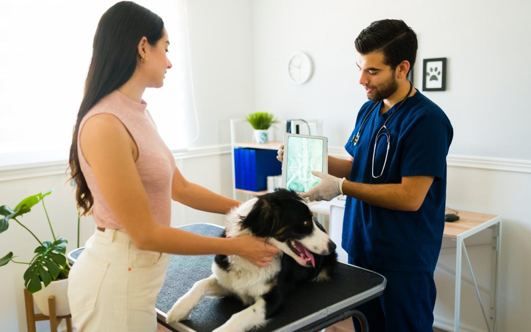
In the ever-evolving field of veterinary medicine, technology plays a crucial role in improving patient care. At our veterinary hospital, we have embraced AI technology—specifically Vetology—for imaging, and the benefits have been remarkable. Let’s explore how this innovative tool has transformed our diagnostic processes and enhanced treatment decisions, ultimately leading to better outcomes for our furry patients.

Team at Avoca Drive Animal Hospital

Meet the team at Avoca Drive Animal Hospital, a Fear Free certified Practice

Streamlining Workflow for Efficiency

From the moment we integrated Vetology into our practice, we focused on making the transition as seamless as possible. Collaborating with our Daysmart software team, we completed the integration within just a couple of days. This investment in time and effort has proven invaluable.

Now, AI-generated reports flow directly into our practice management software, as well as the Vetology workstation platform. This integration eliminates the cumbersome process of manually uploading and downloading files, which previously consumed precious time that our team needed to dedicate to patients and clients. By streamlining our workflow, we can focus more on providing exceptional care.

Enhanced Diagnostic Confidence

Once AI results are incorporated into a patient’s file, our veterinarians leverage this data alongside their expertise. Each case prompts our team to critically analyze the findings:

“Does this make sense given the patient’s history, signalment, and other information that the AI doesn’t have?”

With a comprehensive view that includes history, blood results, and AI insights, our veterinarians gain a clearer understanding of the patient’s condition. This is especially crucial in urgent cases, such as those involving abdominal pain or suspected gastrointestinal issues.

The added assurance that AI provides helps our team confirm that nothing has been overlooked, reducing stress during busy times when multiple cases demand attention.

Making Timely Decisions for Critical Care

When our vets have a clear plan of action, informed by both their clinical judgment and AI reports, we are much better positioned to act swiftly. In urgent situations, this can mean the difference between life and death for seriously ill patients. Rather than waiting for specialist opinions or navigating uncertainties, we can initiate the necessary procedures immediately.

This rapid response allows us to explain diagnoses and obtain consent from pet owners much more efficiently. When every second counts, having reliable AI insights empowers our team to make informed decisions and provide the timely care that pets need.

Conclusion: A New Era in Veterinary Medicine

 The integration of AI technology in our veterinary hospital has transformed the way we approach diagnostics and treatment. By streamlining our processes, enhancing diagnostic confidence, and facilitating timely decision-making, we are better equipped to provide exceptional care for our patients.

As we continue to embrace innovative technologies like Vetology, we remain committed to improving the health and well-being of the pets we serve. The future of veterinary medicine is bright, and we are excited to lead the way in providing the highest standard of care.

Fresh and Refreshed Canine Abdomen Classifiers Now Out

Fresh and Refreshed Canine Abdomen Classifiers Now Out

Screening for Canine Abdomen Conditions Just Got Better with our New and Updated Classifiers

At Vetology, we’re constantly pushing the boundaries of veterinary AI to support your diagnostic needs. That’s why we’re excited to announce the latest update to our Canine Abdomen Virtual AI Radiology Reports!

This release features new and improved classifiers, a combination of the feedback from our clients on our existing canine abdomen classifiers, paired with an expanded range of conditions. Whether you’re an existing client or you’re curious about what Vetology AI can offer, these updates represent a leap forward in diagnostic support for your practice.

Outline drawing of a dog with the abdomen highlighted

What's New?

Improved Performance on Existing Classifiers

Our current canine abdomen classifiers have undergone rigorous retraining and validation to enhance their accuracy. Here’s how we’ve made them better:

  • User Feedback in Action: Direct input through our simple feedback system has been instrumental in guiding our efforts. Coupled with expanded datasets, this process has driven measurable improvements in our AI’s performance.
  • “Golden Set” Validation: Each classifier is tested against a gold-standard dataset reviewed by radiologists to ensure accuracy. While our AI is designed to handle real-world variability, clear and well-positioned radiographs consistently yield the best results.
  • Sensitivity and Specificity: We use a confusion matrix to evaluate classifier performance and only release updates that meet our strict internal standards for reliability and precision.
Expanded Capabilities = More Value

Our newly updated classifiers significantly broaden the range of conditions our AI can identify. With more actionable insights in each report, you can expect even greater support in your diagnostic workflow.

Refined Report Style

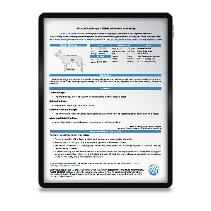
In addition to improved classifiers, we’ve updated the style of our reports to align with the clarity and structure you’ve seen in our recent feline abdomen release. These enhancements provide clearer conclusions and actionable recommendations in a format similar to a board-certified radiologist’s report.

Summary of the conditions and disease processes (classifiers) included in our Canine Abdomen Automated AI Radiology Reports:

Vetology’s AI classifiers are thoughtfully curated to assist veterinarians in addressing complex and frequently encountered conditions requiring radiologist expertise.

The AI-driven screening results in our Virtual AI radiologist report focus on identifying and characterizing diseases that challenge clinical assessment.

  • Liver: Hepatomegaly, hepatic mass, microhepatia.
  • Spleen: Splenomegaly, splenic mass.
  • Kidneys: Right kidney size, right nephroliths, left kidney size, left nephroliths.
  • Gastrointestinal Tract: Mineral/metal gastric material, gastric contents, pyloric/gastric outflow tract obstruction, gastric rugal folds, small intestine distension (mechanical ileus), Small intestine contents, mineral/metal small intestine material, colon diffuse distension, colon contents, spastic colon wall.
  • Urogenital: Urocystoliths/Urethroliths, Mineralized fetal skeletons/pregnancy.
  • Peritoneum: Decreased serosal detail.

Effortless Updates, Seamless Workflow

Vetology AI is designed to save you time while supporting better patient outcomes. By incorporating the latest advancements in machine learning and listening to your feedback, we’re making diagnostic support more reliable and comprehensive than ever.

If you’re not already a Vetology client, now is the perfect time to explore how our AI and teleradiology solutions can transform your practice.

Schedule Your Demo Today!

Want to see how Vetology AI can alter your diagnostic workflow? Contact us today for a demo of our Virtual AI Radiology Reports and teleradiology services.

We encourage you to share this update with your team and colleagues and join us in advancing veterinary diagnostics together.

Pin It on Pinterest