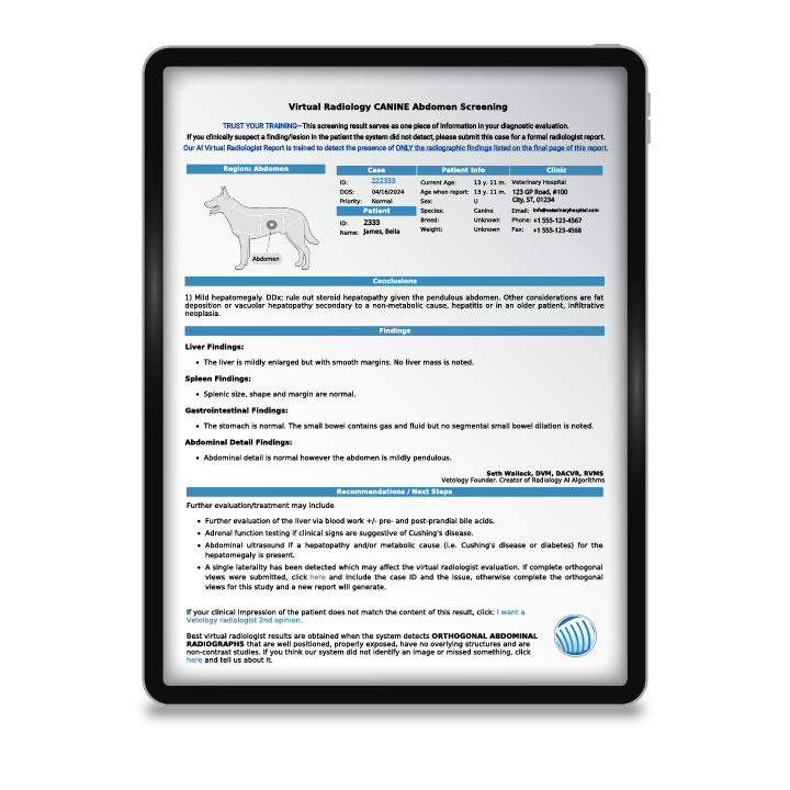
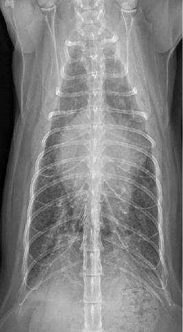
This article examines the comparison between using AI in veterinary radiology and the human experience. Even though AI does improve efficiency by pre-screening X-rays and generating reports, it cannot replace radiologists due to variability in interpretation. AI performs best in clear conditions with strong expert agreement, while complex cases still require human expertise. Read more about how AI in radiology:
- Addresses the shortage of veterinary radiologists.
- Helps with pre-screening and structured reports.
- Works well for conditions like hepatomegaly or pericardial effusion.
- Supports, not replaces, veterinary radiologists.
AI Versus Veterinary Radiologists: Collaboration, Not Competition
About 94 million U.S. households own at least one pet.[1] That’s a lot of furry, feathered, and scaly family members that may potentially need radiographs to diagnose a medical condition. However, there are only 667 board-certified radiologists in the country [2] creating a bottleneck in radiology services. This shortage can correlate to longer wait times, increased anxiety for clinicians and pet owners, and potential delays in diagnosing critical conditions.
This is where artificial intelligence-based radiology tools can help—not to replace veterinary radiologists, but to support them. Artificial intelligence (AI) can pre-screen images, highlight abnormalities, and generate structured reports, allowing radiologists to focus on complicated cases while improving efficiency for general practitioners. But, how does AI compare to human expertise?
Not all conditions are created equal
Radiology is not an exact science but rather an interpretive discipline that relies on pattern recognition, clinical judgement, and experience. Board-certified veterinary radiologists undergo extensive training, but they don’t always agree on image interpretations, especially if the changes are subtle or the patient has multiple diagnoses, creating overlapping signs.
Studies have shown that radiologists tend to have a high level of agreement when interpreting X-rays that display clear and advanced disease. However, variability in interpretation increases when findings are more subtle, as may be the case in early-stage tumors, mild joint changes, or diffuse lung patterns that could indicate interstitial or early inflammatory disease. When subtle abnormalities are suspected, additional imaging, such as ultrasound, computed tomography (CT), or magnetic resonance imaging (MRI), can provide greater anatomical detail and diagnostic confidence.
How interpretive variability affects AI performance assessment
Understanding variabilities in radiologist interpretations is necessary to fairly evaluate the AI’s diagnostic accuracy, sensitivity, and specificity.
- AI algorithms rely on human-labeled data (i.e., ground truth) to learn how to detect and classify abnormalities, and if radiologists don’t agree on a diagnosis, the ground truth may have some degree of subjectivity.
- AI radiology tools are evaluated using accuracy, sensitivity, and specificity, but these measures must be analyzed in the context of how consistently radiologists themselves diagnose the condition.
- If two radiologists interpret the same case differently, the AI may match one but disagree with the other. This doesn’t mean that the AI is wrong; it only highlights the inherent variability in radiology.
How interpretive variability affects AI radiology use
For example, conditions such as hepatomegaly, esophageal enlargement, and the presence of pericardial effusion have a high radiologist agreement rate and are well-suited for AI screening.
At Vetology, each AI-generated report includes a clear list of the conditions assessed, so it’s clear exactly what was evaluated, what was flagged, and what falls outside the scope of the current screening. This provides veterinarians with a solid understanding of the AI’s capabilities and limitations, enabling them to focus their clinical decisions on conditions that were not screened for, without expecting input on findings beyond the AI’s parameters.

Vetology’s AI tools provide guidance for a wide range of thoracic, abdominal, and musculoskeletal conditions in canine and feline patients, including—but not limited to—the following
Abdominal Classifiers
- Liver enlargement
- Masses that may indicate neoplasia or inflammatory processes
- Splenic changes, commonly linked to systemic or localized disease
- Kidney abnormalities such as mineral deposits, structural size variations that may suggest neoplasia, inflammation, or systemic disease
- Bladder and urethral stones
- Pregnancy detection
- Gastrointestinal tract abnormalities, which may indicate obstruction, motility issues, or other conditions
- Peritoneal fluid accumulation, inflammation, or infection
Thoracic classifiers
- Pulmonary patterns
- Cardiomegaly
- Pleural fissure lines
- Fluid accumulation
- Soft tissue pulmonary nodules
- Masses
- Vascular enlargement
Leveraging AI screening alongside teleradiology
Vetology allows veterinarians to optimize AI radiology screening tools and teleradiology services to enhance diagnostic accuracy, improve efficiency, and expedite patient care.
For example, let’s say you handle 60 X-ray cases a month, and you send out only 10 for teleradiologist review to avoid the expense. A Vetology subscription, which provides unlimited access to AI screening and full reports in as little as five minutes, could support your clinical expertise, helping to confirm your suspicions and streamline decision-making. If you still have doubts about a case, you can escalate it for review by a board-certified veterinary radiologist.
This approach creates a three-tiered approach to patient care, integrating:
• AI insights
• With your professional judgement,
• and expert validation from a radiologist when needed.
Collaborating with the Vetology team can help ensure that your patients receive a timely diagnosis and treatment plan, allowing them to receive the care they deserve quickly.


How you can support accurate AI screening and faster board certified radiologist reports
One of the most important factors that lead to an accurate AI screening is good radiographic technique. Clear, well-positioned, well-developed radiographs are necessary for accurate human and AI interpretation, and the AI does not have the ability to adjust its interpretation based on altered positioning or an unclear image.
For example, if a patient is slightly twisted, anatomical structures may appear distorted on the image. This can lead the AI to misread the size or shape of an organ, or even misidentify a condition. Human radiologists can identify when a patient isn’t perfectly positioned and adjust their interpretation, but AI doesn’t yet have that context—it reads exactly what’s in front of it.
You can take the following measures to increase the likelihood of accurate AI screening:
- Ensure proper positioning of each patient
- Choose the correct radiographic settings to ensure a clear image
- Take at least two views (ventrodorsal and lateral views) of the area to be assessed every time.
- Collimate down to the region of interest to reduce scatter.
Vetology offers personalized, on-demand support tailored to answer your needs and questions. Our team of radiologists and veterinary technicians is always available to provide free, one-on-one guidance with positioning skills and technical assistance (in some cases), whether you’re a seasoned practitioner, a new team member, or a recent graduate.
References
[2] AVMA published statistics – veterinary specialists in the United States as of December 31, 2024.AVMA published statistics – veterinary specialists in the United States as of December 31, 2024.

